Cheryl’s Paper Published in Cell Reports Methods! New Stereo-Tracking Method and Discovery of Axon Bubbles

Axon bubbles: hidden structures in the developing brain, seen for the first time!

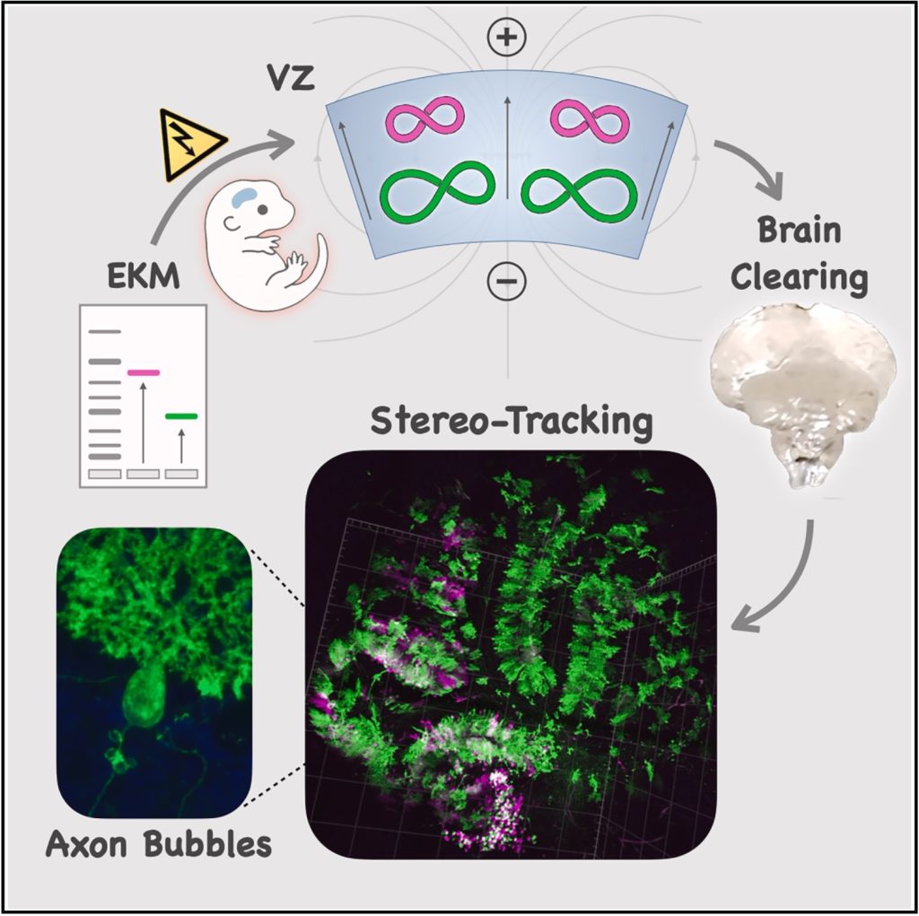
A new method we call Stereo-Tracking takes advantage of a bioelectrical interaction between DNA and cells in the embryonic brain to label the brain’s progenitors in 3D! Combined with brain clearing and light sheet imaging, it lets us track how different neural stem cell niches map onto brain circuits in ways we could not before.

Applying it to the cerebellum, the “little brain” at the back of the head, we stumbled upon a previously unknown cellular structure. We noticed bizarre membranous spheres clustering around the base of growing axons in the second postnatal week of development. They appear completely empty inside, making them invisible to previous labeling methods.

We named these mysterious structures “electrolucent axon bubbles“. Fun fact, we first confirmed their existence on the day of the spring equinox, when they appeared as radiant disks on electron micrographs; hence electrolucent, a small dedication to Apollo, the sun god 🌞. True science anecdote!

These bubbles form on Purkinje cell axons as granule cells squeeze past them during the largest neuronal migration in the brain (don’t forget: the cerebellum contains many more neurons than the rest of the brain combined)!

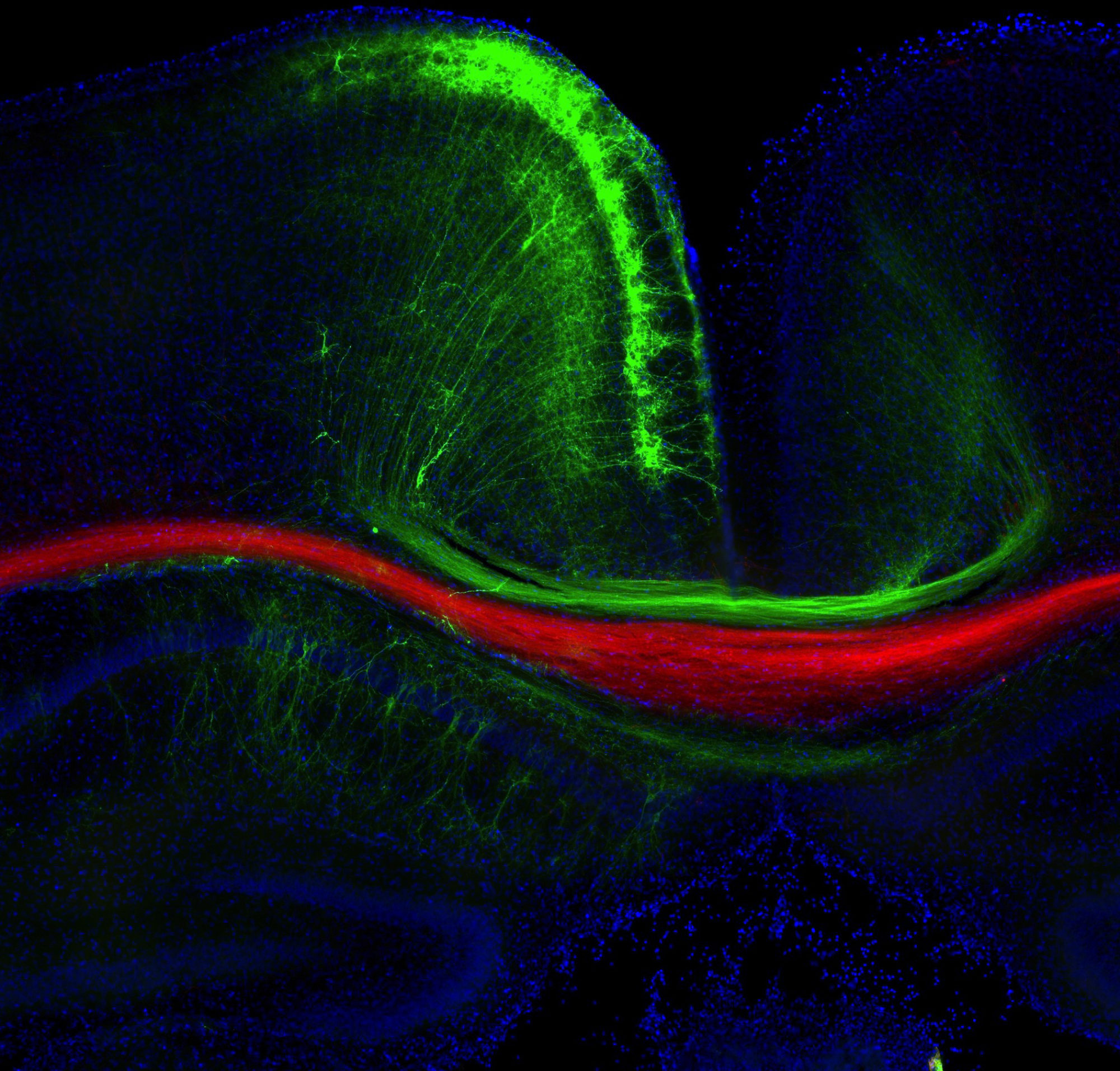
The light sheet rendering below shows Stereo-Tracked Purkinjes as they wire the developing cerebellum. Their striking circuitry quietly handles enormous computational workloads, like GPU arrays running alongside the cerebral cortex’s CPU. Kudos to Dr. Cheryl Brandenburg for these beautiful data!

Read the full story in our paper just out in Cell Reports Methods.

Electrokinetic mobility cerebellum bubbles

Huge congratulations to lead author Cheryl Brandenburg, the entire PouLab team at UMSOM, and our fantastic collaborator teams Roy Sillitoe at Baylor, Ben Cooper at the Max Planck Institute for Multidisciplinary Sciences, and Izumi Sugihara at Tokyo Medical and Dental University!

Big thanks as well to the NIH Common Fund, the Autism Research Institute, and the MPRC Training Program. Without your support, none of this would have been possible!

Colin’s Prime editing in neurons paper out in Methods Mol Biol!

Congrats to Colin Robertson and other lab members for publishing “Prime Editing of Mouse Primary Neurons” on how to use Prime editors on neurons to introduce precise genome edits, including pathological variants from patients.

This paper also includes a step-by-step guide on how to use our web app for pegRNA design and cloning PegAssist.app!

We should be hearing more exciting news from Colin and this powerful new technology for neural somatic genome editing soon…!

Neuroligin-3 paper gets the cover of Biological Psychiatry!

Biol Psychiatry cover, Nov 2024

Congrats to Bek Altas and other lab members, as well as our collaborators at the Max Planck Institute for Multidisciplinary Sciences and the University of Turin. Our paper on the synaptic localization of Neuroligin-3 in the mouse and human brain and the molecular mechanism that regulates it, became the cover article on this month’s issue of Biological Psychiatry! Congrats to Cheryl Brandenburg for the cover art and the epic experiment that inspired it, one of the first ever immunolabelings of excitatory and inhibitory synapses in the human brain!

Neuroligin-3 paper out in Biological Psychiatry!

Just out, our paper on the biology of a synaptic adhesion molecule critically implicated in autism, published in Biological Psychiatry!

This massive paper includes outstanding work from scientists and collaborators at the Max Planck Institute for Multidisciplinary Sciences in Germany, and the University of Turin in Italy. It includes some of the first ever imaging of human synapses (spot the cool cup-shaped presynaptic terminals nestled in the human brainstem ????!), and identifies a molecular mechanism that determines the synaptic localization and transmitter-specificity of Neuroligin-3 between excitatory and inhibitory synapses in the brain. Congrats to everyone involved, a great way to end the year!

Region-Specific Phosphorylation Determines Neuroligin-3 Localization to Excitatory versus Inhibitory Synapses


In utero prime editing of epilepsy variant on bioRxiv!

Modeling an ultra-rare epilepsy variant in wildtype mice with in utero prime editing

For all genome editing fans out there, check out our latest study on bioRxiv showcasing in utero prime editing to model an epilepsy patient with an ultra-rare GRIN variant. To our knowledge, this marks a significant milestone as the first prime editing of neurons in vivo!

Led by Colin Robertson in our lab, alongside Patrick Davis from Boston Children’s Hospital and Harvard Medical School, this ambitious project aims to make precision medicine more accessible to a broad patient population. This rapid workflow to generate personalized animal models with prime editing we hope is a step toward enabling individuals with rare genetic epilepsies to test and tailor pharmacotherapy on their personalized models, reducing the burdens of exploring treatments.

A big thank you for the collaborative efforts of the team, including our partner UM-MIND labs of Peter Crino, Phil Iffland, Steffen Wolff, Brian Mathur, and Ivy Dick; as well as our wonderful collaborators from U Pitt, Eldin Jašarević, and UC Anschutz, Tracy Bale! Thank you all for this achievement and the fantastic collaboration we enjoy!

We look forward to this research advancing toward bedside-to-bench applications!

Bek’s paper out in Journal of Cell Biology!

Congrats to Dr. Bek Altas and our collaborator Prof. Hiroshi Kawabe from Gunma University, Japan, who teased apart a complicated network of cellular interactions between neurons and glia in the brain that cause seizures. Mechanisms of proteostasis and downstream ion gradient homeostasis in astrocytes results in epileptiform network activity in neurons, possibly informing about the pathophysiology behind rare forms of epilepsy with gene variants of the ubiquitin pathway. Read all about it!

Nedd4-2-dependent regulation of astrocytic Kir4.1 and Connexin43 controls neuronal network activity

Cas9-RC on the cover of The CRISPR Journal!

Ryan Richardson’s paper describing the development of Cas9-RC, a new CRISPR agent with increased performance for knockin, is out in the October 2023 issue of The CRISPR Journal.

Not only that, but the paper got the cover! Kudos to Cheryl Brandenburg for the beautiful image of knockin neurons and astrocytes using Cas9-RC in the developing mouse brain. Congrats to the whole team for concluding this large piece of Synth Bio meets Dev Neurosci!

We would be delighted to have you try out Cas9-RC for your own knockin needs. Plasmids will become available on Addgene within the next few days!

Cas9-RC Knockin on the cover of The CRISPR Journal

Phosphorylation of Neuroligin-3

Our work on how the synaptic adhesion molecule Neuroligin-3 is targeted to either excitatory or inhibitory synapses based on phosphorylation is now available on the bioRxiv! Congrats to Bekir Altas, Liam Tuffy, Annarita Patrizi and the rest of the team in this international collaboration between the University of Maryland School of Medicine, the Max Planck Institute for Multidisciplinary Sciences, and the University of Turin.

Phosphorylation Determines Whether Neuroligin-3 is at Excitatory or Inhibitory Synapses in Different Regions of the Brain